 Datasets
 Models
Downloads: 1
Data:
 De-identified
Image
Tabular
Specialty:
De-identified
Image
Tabular
Specialty:
 Oncology
Radiology
Medical Imaging Technique:
Oncology
Radiology
Medical Imaging Technique:
 CT
Medical Imaging Region:
CT
Medical Imaging Region:
 Chest
Omics:
Chest
Omics:
 Transcriptomics
Task:
Transcriptomics
Task:
 Biomarker Discovery
Quantification/Radiomics
License:
Biomarker Discovery
Quantification/Radiomics
License:
 Creative Commons Attribution 3.0
Switch to unified view
| a/README.md | b/README.md | ||
|---|---|---|---|
... |
... |
||
| 8 | 8 | ||
| 9 | For scientific inquiries about this dataset, please contact Dr. Hugo Aerts of the Dana-Farber Cancer Institute / Harvard Medical School (hugo_aerts@dfci.harvard.edu). |
9 | For scientific inquiries about this dataset, please contact Dr. Hugo Aerts of the Dana-Farber Cancer Institute / Harvard Medical School (hugo_aerts@dfci.harvard.edu). |
| 10 | 10 | ||
| 11 | Gene-expression Data Corresponding microarray data acquired for the imaging samples are available at National Center for Biotechnology Information (NCBI) Gene Expression Omnibus (Link to GEO: http://www.ncbi.nlm.nih.gov/geo/query/acc.cgi?acc=GSE58661). The patient names used to identify the cases on GEO are identical to those used in the DICOM files on TCIA and in the clinical data spreadsheet. Clinical Data Corresponding clinical data can be found here: Lung3.metadata.xls. Please note that survival time is measured in days from start of treatment. DICOM patients names are identical in TCIA and clinical data file. |
11 | Gene-expression Data Corresponding microarray data acquired for the imaging samples are available at National Center for Biotechnology Information (NCBI) Gene Expression Omnibus (Link to GEO: http://www.ncbi.nlm.nih.gov/geo/query/acc.cgi?acc=GSE58661). The patient names used to identify the cases on GEO are identical to those used in the DICOM files on TCIA and in the clinical data spreadsheet. Clinical Data Corresponding clinical data can be found here: Lung3.metadata.xls. Please note that survival time is measured in days from start of treatment. DICOM patients names are identical in TCIA and clinical data file. |
| 12 | 12 | ||
| 13 | 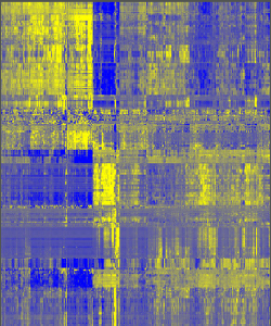 |
13 |  |
| 14 | 14 | ||
| 15 | Publications |
15 | Publications |
| 16 | Aerts, H. J. W. L., Velazquez, E. R., Leijenaar, R. T. H., Parmar, C., Grossmann, P., Cavalho, S., … Lambin, P. (2014, June 3). Decoding tumour phenotype by noninvasive imaging using a quantitative radiomics approach. Nature Communications. Nature Publishing Group. http://doi.org/10.1038/ncomms5006 |
16 | Aerts, H. J. W. L., Velazquez, E. R., Leijenaar, R. T. H., Parmar, C., Grossmann, P., Cavalho, S., … Lambin, P. (2014, June 3). Decoding tumour phenotype by noninvasive imaging using a quantitative radiomics approach. Nature Communications. Nature Publishing Group. http://doi.org/10.1038/ncomms5006 |